-
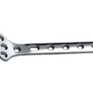
Application:Fractures of the distal radius (applied to the volar aspect). Width: 10mm Thickness: 1.5mm
Ulna & Radius Volar Distal Radius Plate (Small)
-
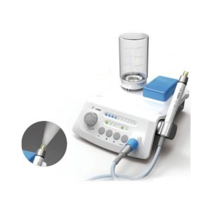
Ultrasonic scaler Features/Specification: VRN-A8 Wireless control scaler with auto-water supply Function: Scaling, Perio, Endo irrigation Water supply: Auto-water supply Wireless control foot switch Handpiece: Metal Detachable handpiece Color: White Input: AC100~240V 47~63Hz Main unit input: DC30V 1.5A Foot switch battery: 1.5V× 2 Receiving sensitivity: -114Db Power: 30VA~48VA Vibration frequency of the tip: 25~31KHz Primary vibration excursion of the tip: ≤ 100μm Output half-excursion: < 2N Ultrasonic output power: 3W~20W Water pressure: 0.01MPa~0.05MPa Weight of main unit: 1.10kg Weight of adapter: 0.25kg
Ultrasonic scaler
-
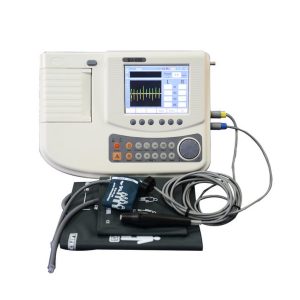
Feature: 1. Obtains two pressures at each ankle site (PVR and DOPPLER) 2. Automated segmental studies to customize the item 3. Automatic ABI, TBI and segmental calculation 4. Performs the Seated ABI on mobility impaired patients 5. Bi-Directional Doppler probe 6. PVR waveform modality & PPG probe to obtain toe and limb pressures Specification Ankle-brachial index(ABI),toe-Brachial Index(TBI) ,segment exams, automatic calculation of the indices Control mode: automatic cuff inflation/deflation and hand-held controller Wave mode: bi-directional doppler, pulse volume recording(PVR), photoplethysmography(PPG) Doppler specification: bi-directional continuous wave(CW), waveform amplitude accuracy:+/- 10%, Probe: 8MHz PPG specification: wave length:940nm, synchronous demodulation, AC paired PVR specification: bandwidth 0.16-12.5Hz, AC paried Display: 5.6inch, TFT, (640*RGB*480) Printer: paper width:80mm, printing width:70mm, resolution 8 points/mm Data storage and transmission: maximum store 100 patient’s data, transmission to computer via USB 2.0 Power supply:100-240VAC, 50-60Hz
Ultrasonic Vascular Doppler Detector
-
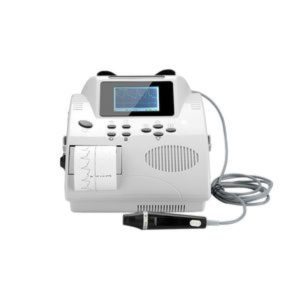
Feature: 620VP TFT LCD displays the wave of the instant blood flow average velocity and strength. It prints the monitoring blood flow curve simultaneously; Detect the blood stream status of arterial/venous by 8MHz probe; Detect the blood flow average velocity, detect the result of fingers/toes and part of body’s vein anatomies operation; BV-620VP(TFT) portable bidirectional vascular Doppler with large color LCD screen; Built-in ARM microprocessor & real-time displays blood flow velocity waveform; Can store 50 detected waveforms; High-speed USB/RS -232 port, can be connected directly to computer to analyze, store, and print the waveform data; Detect peak and average blood speed Detect blood flow of peripheral vessel Detect subsection systolic pressure Detect phlebostenosis, vein occlusions Detect Systolic pressure of toes and fingers Detect blood speed during recovery Detect the pulse rate and show PR simultaneously Specification: Frequency range:Main unit:200±80~5000±1000HZ Probe:350±80~2500±500HZ External output:loudspeaker, single channel with earphone jack Emissive waveform: sine wave Ultrasonic frequency:5.0MHz±10% 、8.0MHz ±10% Ultrasonic frequency average intensity: 100dB Printing speed:40cm/min,60cm/min ,display synchronize with print Frequency mode display range:0.2 KHz~7.0 KHz LCD Display:128 X 64 dots, LCD cursor indicates blood stream speed, double display mode for the spectrum and velocity. Power indication: blue LED indication Charge indication: the charge indicator is yellow when charging and become green after full charge Alarm indication: the indicator light is red when the lack of battery Lack of paper indication: When the recorder is lack of paper, the indication light is red Outline dimension:210*220*105 (mm) Net weight:1.9 Kg Environment condition: Temperature:+5℃ ~ +40℃ , Humidity:<80%, Atmospheric pressure:86kPa ~ 106kPa. Transport and storage environment: Temperature:-10℃ ~ +55℃ , Humidity:<80%, Atmospheric pressure:86kPa ~ 106kPa
Ultrasonic Vascular Doppler Detector Bi
-
SaleProvides high conductive effect and clear, accurate recording without electrical artifacts. Has high resistance to drying (high viscosity). Provides proper adhesion for electrodes. Chemically neutral (no interactions with electrode or skin of patient). Hypoallergenic.
Ultrasound Gel 5Ltr.
Original price was: ₦7,500.00.₦6,987.50Current price is: ₦6,987.50. -
Sale
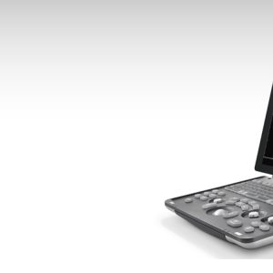
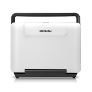
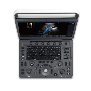
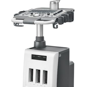
Ultrasound Machine – Sonoscape E2 – Color Ultrasound Scanner (Table Top)
Original price was: ₦12,800,000.00.₦12,500,000.00Current price is: ₦12,500,000.00.Original price was: ₦12,800,000.00.₦12,500,000.00Current price is: ₦12,500,000.00. Add to cartQuick View -
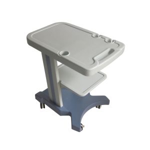
: P/N:BJT-8203-00 Desktop size suitable for all portable ultrasound systems Made of ABS, Strong & Lightweight, easy to clean. Full protection to an ultrasound machine, probes, video printer, etc Built-in Handle on the desktop Built-in holders for various probes and gel Built-in drawer for recorder Trolley size: 680mm L * 450mm W * 750mm H
Ultrasound Trolley BJT-8203-00
-
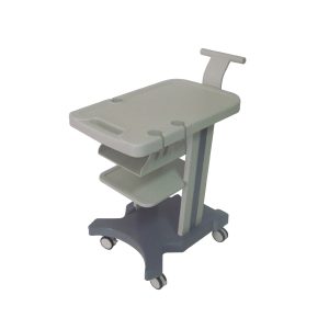
: P/N:BJT-8203-01 Desktop size suitable for all portable ultrasound system Made of ABS, Strong & Lightweight, easy to clean. Full protection to the ultrasound machine, probes, video printer, etc Built-in holders for various probes and gel Handle design for easy transportation Special designed drawer for recorder Trolley size: 680mm L * 450mm W * 750mm H
Ultrasound Trolley BJT-8203-01

Zenrox Healthcare Solutions - Advanced Medical Equipment in Lagos
Zenrox offers cutting-edge medical equipment and comprehensive support services in Lagos. Explore our products designed for superior healthcare delivery. Contact us today for innovative medical solutions.

