-
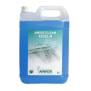 Ingredients Didecyldimethylammonium chloride, chlorhexidine digluconate, non-ionic surfactants, perfume, colouring, excipients. Storage Dangerous – respect the precautions for use (drawn up according to the European rules in force regarding the classification and labelling of chemical products). Storage: between +5°C and +35°C. Products characteristics Detergent and disinfectant designed to reduce the risk of surface biofilm formation on medical devices Broad antimicrobial spectrum to limit the risk of cross-contamination and ensure the safety of personnel Active against MdRB Formulation active in fresh and hard water Formulation non-corrosive and compatible with a broad range of materials Improvement of the ecotoxicological prole: GHG* emissions reduced by 47% relative to a similar product Good rinseability Can be used in an ultrasound cabinet Clear blue solution Pure product pH: min. 6.5 – max. 7.5 pH at 0.5% in mains water: 7.4 Lemon fragrance
Anios Clean Excel D 5 L
-
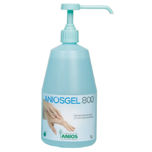 Laboratoires Anios™ Aniosgel 800 Sanitizer is suitable for hygienic treatment and surgical disinfection of hands by rubbing. High level of observance Tested under dermatological control Adapted to sensitive skin Preserves skin hydration Broad antimicrobial spectrum from 30 sec. Conforms to latest European standards Active against bacteria, mycobacteria, yeasts, molds, viruses
Anios Gel 800 1L
-
 Laboratoires Anios™ Aniosgel 800 Sanitizer is suitable for hygienic treatment and surgical disinfection of hands by rubbing. High level of observance Tested under dermatological control Adapted to sensitive skin Preserves skin hydration Broad antimicrobial spectrum from 30 sec. Conforms to latest European standards Active against bacteria, mycobacteria, yeasts, molds, viruses
Anios Gel 800 500 ml
-
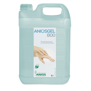 Laboratoires Anios™ Aniosgel 800 Sanitizer is suitable for hygienic treatment and surgical disinfection of hands by rubbing. High level of observance Tested under dermatological control Adapted to sensitive skin Preserves skin hydration Broad antimicrobial spectrum from 30 sec. Conforms to latest European standards Active against bacteria, mycobacteria, yeasts, molds, viruses
Anios Gel 800 5L
-
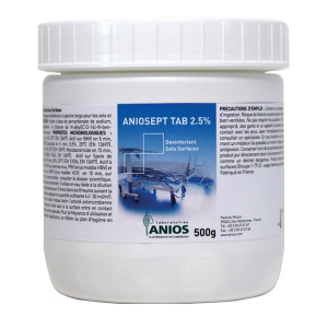 Details Disinfectant product for floors, surfaces and equipment compatible with chlorinated derivatives Quick dissolving effervescent tablet Disinfectant action pH after dissolving in water ~0.5% active chlorine, i.e. 5tablets in 1.5L of water ~6.3 ~0.1% active chlorine, i.e. 2tablets in 3L of water ~6.7
Aniosept Tab 2.5% 500g 150’S
-
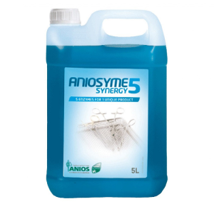 Cleaning of surgical and medical equipment, endoscopes and medical devices by soaking, ultrasonic tank, automatic washing machine and washing tunnel. ANIOSYME SYNERGY 5, with its 5 enzymes (amylase, cellulase, lipase, mannanase and protease), provides a broad spectrum of performance on the constituents of all types of stains.
Aniosyme Synergy 5 5L
-
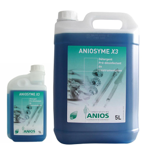 Aniosyme X3 available in 1 Ltr and 5 Ltr Reinforced cleaning and pre-disinfection of medico-surgical instrumentation, medical devices and endoscopic equipment Very high performing detergency Cleaning and degreasing action Clear blue liquid optimised antimicrobial efficacy from 5 minutes Cleaning in ultrasonic bins Collection of contaminated instrumentation Tri-enzyme complex combined with a new generation onionic surfactant Neutral pH : compatible with aluminium alloys Chloride free formula : non corrosive on materials pH of pure product : around 8
Aniosyme X3
-
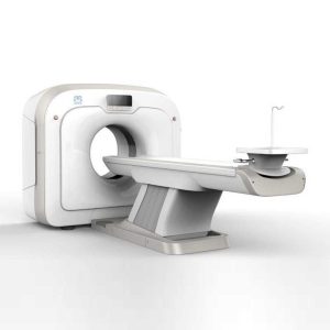
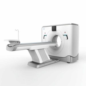
 ANATOM16 HD, is a tool of precision medicine in diagnosis imaging. Via the breakthrough designs in precise hardware, software and imaging technologies, ANATOM 16 HD can provide precise diagnosis information and early detection for small lesions. Features: Seamlessly upgrade to meet your future needs: Anke takes full consideration of the increasing clinical requirements of your business in today’s rapidly changing medical environment. Precise hardware, Precise technology, Precise imaging: OptiWave detector, High precision gantry control, Dual-mode gantry tilt, Admir3D iterative technology, Dual-energy head imaging, 1024 x1024 matrix imaging technology, High-definition imaging of targeted organs, Low dose platform, 3D enhanced VR Precision Technology Platform: ANATOM precision technology platform is equipped with advanced imaging technologies, and adopts OptiWave detector, Ahead dual-energy imaging, Admir3D iterative reconstruction technology and AccuTilt dual-mode tilt gantry technology to provide powerful support for accurate diagnosis Admir3D iterative reconstruction technology: Admir applies mathematical and physics models to accurately construct and describe the signal’s quantum characteristics. Iterative operations are performed in the three domains of raw data, projection and image, to greatly reduce the image noise and achieve optimal image quality with low dose Ahead-Head dual-energy head imaging technology: Ahead creatively uses 140kV and 80kV dual energy switching scan mode for brain imaging. By careful analyzing the high and low energy characteristics, images can show more valuable information about the brain tissues AccuTilt dual-mode gantry tilt technology: The system provides digital and mechanical tilt to accommodate different user habits and clinical needs. Real-time collision preventing system is available for the patients’ safety AccuOrgan-Targeted organ imaging: To achieve high precision imaging of each part of human body at low dose and low energy consumption AccuDose-Comprehensive low dose imaging: Pediatric Scan Protocol, Individual Dose Monitoring, AccuShape Filter, Efficient Detector, Adose Dose Modulation, Ahead – Head Dual-energy Imaging, Iterative Reconstruction, Amast, Contrast Agent Tracking Technology AccuScan-Enjoy ease: Convenient and efficient operation process greatly improve work efficiency to achieve high volume of patients Clinical Applications: Fast, precise and low-dose imaging technologies provide a full range of clinical solutions to meet the current and future clinical diagnostic needs Service Innovation creating maximum value for customers: Service Support within 24 Hours, Local Service Partners, On-line Service Support, After-sales Maintenance Stations AccuSaving Green & Energy-saving: AccuSaving is an innovative energy saving technology. The system will enter the “dormant”, which is a low carbon mode, after a certain idle time or per user’s request. To bring the system back to working status is as easy as pushing a button. The system will also remind the user to perform necessary warm-up and calibration procedures, which are fully automated processes. AccuSaving technology can reduce operation and standby power consumption and save the electricity cost by 30% by adopting different operation modes in working and off hours Technical Specifications: No. Technical feature 1 Gantry 1.01 Gantry type Low voltage slip-ring with AccuSlip-ring technology 1.02 Gantry driven type Strap-driven 1.03 Patient opening 70cm 1.04 Gantry tilt mode Dual-mode gantry tilt 1.05 Mechanical tilt capability ±30° 1.06 Digital tilt capability ±50° 1.07 Gantry remote-Control Provided 1.08 Detector type OptiWave rare-earth ceramic detector 1.09 Numbers of detector rows 32 1.10 Width of Z-axle detector 20mm 1.11 Detector columns of channels per row 912 1.12 Numbers of detector columns 29184 1.13 Data-transfer type RF,optical fiber communication 1.14 3D laser orientation Provided 1.15 External X-ray enable Interface for Foot-Pedal Provided 1.16 Automatic exposure control(mA Modulation) Provided 1.17 Auto-voice manager Breath Graphical Display Hold Message (Record/Playback) Breath Message(Record/Playback) 1.18 ANKE energy conservation management Provided 1.19 Acquisition mode 16 × 0.625mm, 16 × 1.25mm 2 Scan parameter 2.01 Shortest 360 degree rotation time 0.5s 2.02 Allowed rotation times 0.5s,0.8s,1.0s,1.5s,2.0s 2.03 Slice numbers per rotation 16 2.04 Minimum slice thickness of scan 0.625mm 2.05 Minimum slice thickness of reconstruction 0.625mm 2.06 Maximum slice thickness of scan 10mm 2.07 Nominal reconstruction slice thickness 0.625mm,1.25mm,2.5mm,5.0mm, 7.5mm,10mm 2.08 Speed of image reconstruction(512×512) 65 frames/s 2.09 Scan FOV 52cm 2.10 Image reconstruction matrix 512×512,1024×1024 2.11 Image display matrix 512×512,1024×1024 2.12 Maximum continuous scan duration 120s 2.13 Maximum continuous scan length 180cm 2.14 Direction of TOPO Front-back,Left-right 2.15 Max. length of TOPO 180cm 2.16 Range of pitch 0.5~1.5 2.17 Scan mode Scout scan Axial scan Helical scan Cine scan 3 HVPS and Tube 3.01 Maximum continuous output of HV generator 50kW 3.02 Tube kV selections 80kV,100 kV,120 kV,140 kV 3.03 Tube mA range 10~420mA 3.04 Tube anode heat capacity 5.0MHU 3.05 Heat dissipation rate 815kHU/min 3.06 Type of cooling Oil cooling + Air cooling 3.07 Tube focus Large:1.0 mm×1.0mm Small:0.5mm×1.0mm 3.08 Dynamic flying focal spot technology Provided 4 Patient table 4.01 Maximum horizontal-movable range 1850mm 4.02 Table horizontal-scannable range 1800mm 4.03 Table horizontal-position repeatability ±0.25mm 4.04 Maximum vertical-movable range 500mm 4.05 Maximum speed of vertical movement 20mm/s 4.06 Maximum speed of horizontal movement 150mm/s 4.07 Maximum patient weight 205kg 4.08 Foot pedal of patient table control Provided 5 Image Quality 5.01 High contrast resolution 21lp/cm@0%MTF 5.02 Low contrast resolution 2.0mm@0.30% 5.03 Isotropic imaging resolution 0.625mm 5.04 Range of CT numbers -32767~32768 5.05 Image noise ≤0.25@28mGy 6 Computer subsystem 6.01 CPU 3.5GHz 6.02 Memory 16GB×4 6.03 Storage of hard-disk 1T×2 6.04 Monitor 24’’ LCD Monitor 6.05 Resolution of monitor 1920×1200 6.06 Image-data external storage type CD/DVD/USB 6.07 Time of image reconstruction(512×512) 15.4ms/frame 6.08 DICOM 3.0 interface Provided 6.09 Printer DICOM 3.0 interface Provided 6.10 Auto filming Provided 6.11 Worklist function Provided 7 Advanced application 7.01 Multi-Planar Reconstruction(MPR) Provided 7.02 Curve Multi-Planar Reconstruction(CPR) Provided 7.03 Surface Shaded Display(SSD) Provided 7.04 Volume Rendering(VR) Provided 7.05 Maximum Intensity Projection(MIP) Provided 7.06 Minimum Intensity Projection(MinIP) Provided 7.07 Virtual Endoscopy(VE) Provided 7.08 CT angiography(CTA) Provided 7.09 Tissue segmentation Provided 7.10 One click bone remove Provided 7.11 One click patient table remove Provided 7.12 Bolus-tracking Technology Provided 7.13 Spiral auto start Provided 7.14 Cine display Provided 7.15 AbastTM bone artifact suppression technology Provided 7.16 AmastTM metal artifact suppression technology Provided 7.17 Admir3D fulll-domain iterative reconstruction Provided 7.18 Low-dose pediatric scan technology Provided 7.19 Low-dose lung scan technology Provided 7.20 AccuHead grey-white matter enhanced technology Provided 7.21 AccuLung high resolution scan technology Provided 7.22 AccuOtica inner-ear high resolution scan technology Provided 7.23 AccuBody high resolution scan technology Provided 7.24 AccuBone high resolution scan technology Provided Click Here To Download Catalogue
Anke Anatom 16 Slice CT Scan
-
 This Machine gives a possibility to perform computed tomography without any problems and on high quality level. This device is used to conduct exams of internal organs and their functioning. With its help, a physician has a possibility to assess the condition of the human body as a whole. Features: It is easy to use; Convenience; Multi functionality; Obtained images are of high definition; High-definition 3D images of the area under study; The procedure is pain-free; The data is processed fast; The image can be stored in the computer memory; The diagnostics does not take a lot of time; Acceptable radiation dose. Technical Specifications: No. Technical Features s 1 Gantry 1.01 Gantry type Low voltage slip-ring 1.02 Gantry driven type Strap-driven 1.03 Patient opening 70cm 1.04 Gantry tilt mode Digital gantry tilt 1.05 Digital tilt capability ±50° 1.06 Detector type OptiWave rare-earth ceramic detector 1.07 Numbers of detector rows 16 1.08 Width of Z-axle detector 20mm 1.09 Detector columns of channels per row 848 1.10 Numbers of detector columns 13568 1.11 Data-transfer type RF, optical fiber communication 1.12 Distance of focus-ISO-center 53cm 1.13 Distance of focus-detector 94cm 1.14 3D laser orientation Provided 1.15 13″ integrated display panel Provided 1.16 Adose automatic exposure control (mA Modulation) Provided 1.17 Auto-voice manager Breath Graphical Display Hold Message (Record/Playback) Breath Message (Record/Playback) 1.18 AccuSaving energy conservation management Provided 2 HVPS and X-ray tube 2.01 Maximum continuous output of HVgenerator 42kW 2.02 Tube kV selections 70kV, 80kV, 100 kV, 120 kV, 140 kV 2.03 Tube mA range 10~350mA 2.04 Tube anode heat capacity 3.5MHU 2.05 Max. anode cooling rate 735kHU/min 2.06 Type of cooling Oil cooling + Air cooling 2.07 Tube focus Large: 1.2mm×1.4mm Small: 0.7mm×0.8mm 2.08 Collimator width selection 4-level election 2.09 Focus spot tracking technology Provided 3 Patient table 3.01 Maximum horizontal-movable range 1850mm 3.02 Table horizontal-scannablerange 1800mm 3.03 Table horizontal-position repeatability ±0.25mm 3.04 Minimum height above floor 430mm 3.05 Maximum vertical-movable range 500mm 3.06 Maximum speed of vertical movement 35mm 3.07 Maximum speed of horizontal movement 150mm/s 3.08 Maximum patient weight 205kg 3.09 Foot pedal of patient table control Provided 4 Computer 4.01 CPU 3.5GHz 4.02 Memory 32GB 4.03 Storage of hard-disk 1TB×2 4.04 Monitor 24’’ LCD Monitor 4.05 Resolution of monitor 1920×1200 4.06 Image-data external storage type CD/DVD/USB 4.07 Time of image reconstruction (512×512) 33.3ms/image 4.08 Speed of image reconstruction (512×12) 30fps 4.09 DICOM 3.0 interface Provided 4.10 Printer DICOM 3.0 interface Provided 4.11 Auto filming Provided 4.12 Worklist function Provided 5 Scan parameters 5.01 Shortest 360 degree rotation time 0.75s 5.02 Allowed rotation times 0.75s, 1.0s, 1.5s, 2.0s, 3.0s, 4.0s 5.03 Maximum slice numbers per rotation 32 5.04 Minimum slice thickness of scan 1.25mm 5.05 Minimum slice thickness of reconstruction 0.625mm 5.06 Maximum slice thickness of scan 20mm 5.07 Nominal reconstruction slice thickness 0.625mm, 1.25mm, 2.5mm, 5.0mm, 7.5mm, 10mm, 20mm 5.08 Speed of image reconstruction (512×512) 30 frames/s 5.09 Scan FOV 50cm 5.10 Image reconstruction matrix 512×512, 1024×1024 (Optional) 5.11 Image reconstruction matrix 512×512, 1024×1024 (Optional) 5.12 Image display matrix 512×512, 1024×1024 (Optional) 5.13 Maximum continuous scan duration 120s 5.14 Maximum continuous scan length 180cm 5.15 Direction of TOPO Front-back, Left-right 5.16 Max. length of TOPO 180cm 5.17 Range of pitch 0.5~1.5 5.18 Scan mode Scout scan Axial scan Helical scan Cine scan 6 Image Quality 6.01 High contrast resolution 21lp/cm@0%MTF 6.02 Low contrast resolution 2.0mm@0.30% 6.03 Isotropic imaging resolution 0.24mm 6.04 Range of CT numbers -32767~32768 6.05 Image noise ≤0.29@28mGy 7 Advanced application 7.01 Multi-Planar Reconstruction (MPR) Provided 7.02 Curve Multi-Planar Reconstruction (CPR) Provided 7.03 Surface Shaded Display (SSD) Provided 7.04 Volume Rendering (VR) Provided 7.05 Maximum Intensity Projection (MIP) Provided 7.06 Minimum Intensity Projection (MinIP) Provided 7.07 Virtual Endoscopy (VE) Provided 7.08 CT angiography (CTA) Provided 7.09 Tissue segmentation Provided 7.10 One click bone remove Provided 7.11 One click patient table remove Provided 7.12 Bolus-tracking Technology Provided 7.13 Spiral auto start Provided 7.14 Cine display Provided 7.15 AbastTM bone artifact suppression technology Provided 7.16 AmastTM metal artifact suppression technology Provided 7.17 Admir3D all-domain iterative reconstruction Provided 7.18 Low-dose pediatric scan technology Provided 7.19 Low-dose lung scan technology Provided 7.20 AccuHead grey-white matter enhanced technology Provided 7.21 AccuOrgan lung high resolution scan technology Provided 7.22 AccuOrgan inner-ear high resolution scan technology Provided 7.23 AccuOrgan body high resolution scan technology Provided 7.24 AccuOrgan bone high resolution scan technology Provided 7.25 AccuMatter dual-energy with Admir3D for new application Provided Click Here To Download Catalogue
Anke Anatom 32 Fit Multi-Slice Spiral CT Scan
-
 The ANATOM 64 CT scanner is the latest innovation for cardiac imaging based on Precision Platform system. The excellent design of Ahart technology which innovatively combined single spiral scan + gated imaging + mA modulation for easy heart imaging at extremely low radiation dose. We provide you ANATOM 64 Clarity/Precision of two models which are low/high configurations for preferences. It also offers you conventional clinical applications of low dose, better image quality and faster exams. Features: Modularized OptiWave HD detector features low-cost & easy maintenance, high spatial resolution and long lifetime Admir3D iterative technology delivers optimal dose efficiency and noise reduction without compromising image quality High configurations of main components ensure the best results and maximum patient throughput Uniquely and creatively uses 140kV and 80kV dual energy scan mode for brain imaging on 16-slice CT to offers you extraordinary image quality both in low and high density resolutions AdoseTM mA modulation ensures you low dose imaging without compromising image quality particularly useful in pediatric applications Equipped with dedicated Abast and Amast for bone and metal artifacts The brilliant Ahart technology enables you to experience so easy and low-dose cardiac imaging applications Technical Specifications: Model ANATOM 64 Precision ANATOM 64 Fit Rack type Low pressure slip ring Low pressure slip ring Scan aperture 70cm 70cm Rack Physical inclination ± 30 ° N.A Rack digital inclination ± 50 ° ± 50 ° cooling method Air-cooled Air-cooled Focus to the center distance 56 cm 53 cm Maximum power (non-equivalent) 80kW 42kW Votage (kV) 80kV / 100kV / 120kV / 140kV 70kV / 80kV / 100kV / 120kV / 140kV Heat capacity 8MHU 5.0MHU Heat dissipation rate 931 kHU / min 748kHU / min cooling method Oil cool Oil cool Large focus size 1.1mm × 1.2mm 1.2mm × 1.4mm Small focus size 0.6mm × 1.2mm 0.7mm × 0.8mm Tube current range 10-670mA 10-350mA Detector type Optiwave detectors Optiwave detectors Number of Z-axis 32 32 The width of the Z-axis 20mm 20mm The number of elements per row 912 848 Total number of detectors 29184 27136 Acquisition mode 64×0.625, 32×0.625, 16×0.625 64×0.625, 32×0.625, 16×0.625 Scanning range 1800mm 1800mm Horizontal positioning accuracy ± 0.25mm ± 0.25mm weight capacity 205kg 205kg Minimum height 43cm 43cm Anti – collision protection device Yes Yes Foot control switch Yes Yes IV rack Yes Yes CPU 3.5GHz 3.5GHz RAM 16 GB × 4 16 GB × 4 Hard drive capacity 1T × 2 1T × 2 Display size 24 inch LCD monitor 24 inch LCD monitor Display resolution 1920 × 1200 1920 × 1200 Computer operating system Windows 7 Windows 7 Image reconstruction speed 65 frames/ second 65 frames/ second Number of image store 1000000 1000000 Data external storage mode CD / DVD / USB CD / DVD / USB Minimum Scan Time of 360 degree 0.39sec 0.75sec Sub-millimeter acquisition layers 64 64 Double sub-millimeter acquisition layers 64 64 Thinnest acquisition thickness 0.625mm 0.625mm The thinnest reconstruction thickness 0.3125mm 0.625mm Conventional reconstruction thickness (mm) 0.3125 mm, 0.625 mm, 1.25 mm, 2.5 mm, 5.0 mm, 7.5 mm, 10 mm 0.625 mm, 1.25 mm, 2.5 mm, 5.0 mm, 7.5 mm, 10 mm The reconstruction matrix 512 x 512, 1024 x 1024 512 x 512, 1024 x 1024 Display matrix 1024 × 1024 1024 × 1024 Max FOV 52cm 50cm The maximum display field of view 70cm 50cm Maximum scan length 1800mm 1800mm Maximum continuous helix scan time 120s 120s Pitch range 0.5-1.5 0.5, 1.0, 1.5 High contrast resolution 21 Lp / cm @ 0% MTF 21 Lp / cm @ 0% MTF Low contrast resolution 2mm @ 0.3% 2mm @ 0.3% Image noise ≤ 0.25 ≤ 0.29 MPR Yes Yes CPR Yes Yes SSD Yes Yes VR Yes Yes MIP Yes Yes MinIP Yes Yes Virtual endoscopy Yes Yes CT angiography Yes Yes Tissue segmentation Yes Yes One-key bone removal Yes Yes Automatically patient table removal Yes Yes Contrast Agent Automatic Tracking Technology- bolus tracking Yes Yes Automatic linkage trigger technology Yes Yes Cine mode display Yes Yes Bone artifact suppression technique AbastTM AbastTM Metal artifact suppression technique AbastTM AbastTM Iterative reconstruction technique Admir3D global iteration Admir3D full-domain iteration Low – dose children ‘s scanning technology Yes Yes Low – dose lung scan Yes Yes Gray matter enhancement technology AccuHead AccuHead High resolution imaging of the lung AccuLung AccuLung Inner ear high resolution imaging AccuOtica AccuOtica Body high resolution imaging AccuBody AccuBody Bone high resolution imaging AccuBone AccuBone Head dual-energy imaging Ahead Ahead CT perfusion imaging Optional Optional Quantitative analysis of blood vessels Optional Optional Heart coronary artery imaging Aheart N.A ECG gated Yes N.A Low dose cardiac scan Yes N.A Green energy saving technology AccuSaving AccuSaving Dual-energy scan technology Optional Optional Click Here To Download Catalogue
Anke Anatom 64 Clarity Multi-Slice Spiral CT Scan
-
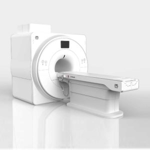
 OPENMARK 5000 is 0.51T MRI. It’s approved by FDA and has CE mark. It adopts two-pillar magnet design with 280 degree openness and equipped with powerful RF and gradient system, together with advanced imaging technology, making it as a high-end system which is comparable to high-field MRI. Features: With the highest system stability and the highest homogeneity of the magnet field in permanent MRI Screens on both sides facilitate positioning; 280 degree two-pillar magnet design ensures stable magnet structure and facilitates interventional treatment. Active and passive shimming calibrate technology ensures the magnetic field uniformity Motor-driven patient couch makes it easier for patients to access and for positioning Powerful hardware and software platforms ensure the scan speed, image quality and make it possible for advanced imaging functions Fast scan speed eliminates motion artifact Rich scan sequences, advanced imaging technology and powerful postprocessing technology ensure image quality, extend more applications, which can fully satisfy the clinical needs Intelligent user-friendly operating system ensures you easy operation Technical Specifications: No. Technique Parameter 1 Magnet System 1.1 Magnet Type Permanent Magnet Automatic constant temperature system 1.2 Field Strength 0.51T 1.3 Magnet Shape Dual-pillar shape 1.4 Homogeneity(40cm,DSV,VRMS) ≤1.6ppm 1.5 Shim Method Active/Passive 1.6 Magnet Vertical Gap (Cover) 40cm 1.7 Magnetic Pole Dia. (Exclude Cover) 145cm 1.8 Accessibility(Horizontal Opening Angle, 280° 1.9 5 Gauss fringe field X-axis:horizontal ≤2.5m Y-axis:Vertical ≤2.5m Z-axis:horizontal ≤2.5m 2 Patient Couch and Communication 2.1 Patient Couch Driven mode Motor-driven 2.2 Max. Patient Weight ≥200kg(440lbs) 2.3 Patient Positioning Tools Laser Light Localizer for positioning of center slice Motor-driven transfer to center of imaging volume 2.4 Position accuracy ±1mm 2.5 Emergency Call Key Yes 2.6 Intercom System Yes 3 Gradient System 3.1 Gradient Field Strength(Single Axis) ≥30mT/m 3.2 Gradient Slew Rate (Single Axis) ≥100mT/m/ms 3.3 Rise Time ≤0.3ms 3.4 Gradient Cooling System ( Gradient coils and Power electronics) Air Cooling 4 RF System 4.1 RF System Type Digital Transmit and Receive signal 4.2 Number of RF Channels 4 4.3 Transmitter Amplifier Peak Power 6kW 4.4 RF Bandwidth of Receiver ≥1.25MHz 4.5 Head Coil Yes 4.6 Neck Coil Yes 4.7 Body/Spine Coil (17 inch) Yes 4.8 Body/Spine Coil (21 inch) Yes 4.9 Knee Coil Yes 4.10 Shoulder Coil Yes 4.11 Flexible Coil Optional 4.12 Breast Coil Optional 5 Computer System 5.1 Host Computer DELL Computer (for MR) 5.2 System Software Windows XP 5.3 Operation Software APEX 5.4 CPU Clock rate 3.0GHz 5.5 Main Memory 4GB 5.6 Color LCD Monitor 19” 5.7 Keyboard and Mouse Standard 5.8 Image Reconstruction Speed(256 x 256 Matrix) 200 frame/Sec. 5.9 Hard Disk 500GB 5.10 Image Storage Capacity(256 x 256 Matrix) 500,000 5.11 Media Driver DVD RW 5.12 DICOM 3.0 Yes 5.13 Ethernet Yes 5.14 Operation Console Yes 5.15 Operation Chair Yes 6 Scanning Parameter 6.1 Max. FOV 410mm 6.2 Min. FOV 5mm 6.3 Min. TE(SE) 5ms 6.4 Min. TR(SE) 11ms 6.5 Min. TE(GR) 1ms 6.6 Min. TR(GR) 3ms 6.7 Min. 2D Thickness 1.0mm 6.8 Min. 3D Thickness 0.1mm 6.9 Max. Image Matrix 512×512 7 Scanning Sequence & Imaging Technique 7.1 Spin Echo 2D/3D (SE 2D/3D) Yes 7.2 DE/QE Yes 7.3 Fast Spin Echo 2D/3D(FSE 2D/3D) Yes 7.4 Single Shot FSE 2D/3D Yes 7.5 Inversion Recovery(IR) Yes 7.6 Fast Inversion Recovery(FIR) Yes 7.7 Gradient Echo 2D/3D(GR 2D/3D) Yes 7.8 Fast GR 2D/3D Yes 7.9 SPGR Yes 7.10 FLAIR Yes 7.11 Fat Imaging Yes 7.12 Fat Suppression imaging Yes 7.13 Water-Fat Separation imaging Yes 7.14 TOF MRA(2D/3D) Yes 7.15 MRCP(2D/3D) Yes 7.16 MRU (2D/3D) Yes 7.17 MRM Yes 7.18 Fast Hydrograph Imaging Yes 7.19 Diffusion Weighted Imaging(DWI) Yes 7.20 Max. b Value 1000s/mm2 7.21 Breath Hold Technology Yes 7.22 Magnetization Transfer Contrast(MTC) Yes 7.23 Multi-slice and Angle-free Presaturation Yes 7.24 Saturation Tracking Yes 7.25 Maximum Intensity Projection(MIP) Yes 7.26 Multi-Angle Projection(MAP) Yes 7.27 3D Reconstruction Yes 7.28 Multi-planar Reconstruction(MPR) Yes 7.29 Multi-Artifacts Eliminating technology Yes 7.30 Checking with Part Metal Implant Yes 7.31 Online Image Filtration Yes 7.32 Online Post Procession Yes 7.33 3D Scout Yes 7.34 Scanning Protocol Preset Yes 7.35 Scanning Protocol Queue Waiting Yes 7.36 Advanced Image Post Processing Yes 7.37 Image Fusion Technology of Vascular Yes 7.38 Image Fusion Technology of Spine Yes Click Here To Download Catalogue
Anke MRI Openmark 5000 Permanent System
-
 SuperMark 1.5T is a new generation superconducting MRI system based on years of experience in production and research. It’s applicable to whole body scan, such as, nervous system, spine, joint soft tissue, pelvic and abdominal cavity, etc. SuperMark 1.5T provides not only conventional pulse sequences and clinical diagnosis functions, but also provides advanced functional applications, for instance, 3D angiography and water imaging. It adopts brand new ANKE APEX operating system which ensures easy operation and fast diagnosis. Technical Advantages: Reliable short cavity superconducting magnet system with zero liquid helium consumption New generation fully digitalized and extensible multichannel spectrometer Powerful high efficiency and high fidelity gradient system; Multi-channel PA RF receiving coil with intelligent identification English operating system and high extensible computer system High resolution conventional clinical images; Practical advanced functional imaging Superconducting MRI System: Highly open and humanization design -> Streamlined design Rich sequences and technology satisfy clinical needs -> Efficient service Low Investment: High cost performance superconducting MRI system Zero liquid helium consumption, low running and maintenance cost Core technology by independent R & D supports full upgrade Low electric consumption Compact magnet design, minimum installation space: 35 square meters High Return: High resolution thin slice images improve diagnosis Short cavity magnet design makes patients comfortable Fast scan speed improves work efficiency Technical Specifications: No. Technique Parameter 1 Magnet System 1.1 Magnet Type Permanent Magnet Automatic constant temperature system 1.2 Field Strength 0.51T 1.3 Magnet Shape Dual-pillar shape 1.4 Homogeneity(40cm,DSV,VRMS) ≤1.6ppm 1.5 Shim Method Active/Passive 1.6 Magnet Vertical Gap (Cover) 40cm 1.7 Magnetic Pole Dia. (Exclude Cover) 145cm 1.8 Accessibility(Horizontal Opening Angle, 280° 1.9 5 Gauss fringe field X-axis:horizontal ≤2.5m Y-axis:Vertical ≤2.5m Z-axis:horizontal ≤2.5m 2 Patient Couch and Communication 2.1 Patient Couch Driven mode Motor-driven 2.2 Max. Patient Weight ≥200kg(440lbs) 2.3 Patient Positioning Tools Laser Light Localizer for positioning of center slice Motor-driven transfer to center of imaging volume 2.4 Position accuracy ±1mm 2.5 Emergency Call Key Yes 2.6 Intercom System Yes 3 Gradient System 3.1 Gradient Field Strength(Single Axis) ≥30mT/m 3.2 Gradient Slew Rate (Single Axis) ≥100mT/m/ms 3.3 Rise Time ≤0.3ms 3.4 Gradient Cooling System ( Gradient coils and Power electronics) Air Cooling 4 RF System 4.1 RF System Type Digital Transmit and Receive signal 4.2 Number of RF Channels 4 4.3 Transmitter Amplifier Peak Power 6kW 4.4 RF Bandwidth of Receiver ≥1.25MHz 4.5 Head Coil Yes 4.6 Neck Coil Yes 4.7 Body/Spine Coil (17 inch) Yes 4.8 Body/Spine Coil (21 inch) Yes 4.9 Knee Coil Yes 4.10 Shoulder Coil Yes 4.11 Flexible Coil Optional 4.12 Breast Coil Optional 5 Computer System 5.1 Host Computer DELL Computer (for MR) 5.2 System Software Windows XP 5.3 Operation Software APEX 5.4 CPU Clock rate 3.0GHz 5.5 Main Memory 4GB 5.6 Color LCD Monitor 19” 5.7 Keyboard and Mouse Standard 5.8 Image Reconstruction Speed(256 x 256 Matrix) 200 frame/Sec. 5.9 Hard Disk 500GB 5.10 Image Storage Capacity(256 x 256 Matrix) 500,000 5.11 Media Driver DVD RW 5.12 DICOM 3.0 Yes 5.13 Ethernet Yes 5.14 Operation Console Yes 5.15 Operation Chair Yes 6 Scanning Parameter 6.1 Max. FOV 410mm 6.2 Min. FOV 5mm 6.3 Min. TE(SE) 5ms 6.4 Min. TR(SE) 11ms 6.5 Min. TE(GR) 1ms 6.6 Min. TR(GR) 3ms 6.7 Min. 2D Thickness 1.0mm 6.8 Min. 3D Thickness 0.1mm 6.9 Max. Image Matrix 512×512 7 Scanning Sequence & Imaging Technique 7.1 Spin Echo 2D/3D (SE 2D/3D) Yes 7.2 DE/QE Yes 7.3 Fast Spin Echo 2D/3D(FSE 2D/3D) Yes 7.4 Single Shot FSE 2D/3D Yes 7.5 Inversion Recovery(IR) Yes 7.6 Fast Inversion Recovery(FIR) Yes 7.7 Gradient Echo 2D/3D(GR 2D/3D) Yes 7.8 Fast GR 2D/3D Yes 7.9 SPGR Yes 7.10 FLAIR Yes 7.11 Fat Imaging Yes 7.12 Fat Suppression imaging Yes 7.13 Water-Fat Separation imaging Yes 7.14 TOF MRA(2D/3D) Yes 7.15 MRCP(2D/3D) Yes 7.16 MRU (2D/3D) Yes 7.17 MRM Yes 7.18 Fast Hydrograph Imaging Yes 7.19 Diffusion Weighted Imaging(DWI) Yes 7.20 Max. b Value 1000s/mm2 7.21 Breath Hold Technology Yes 7.22 Magnetization Transfer Contrast(MTC) Yes 7.23 Multi-slice and Angle-free Presaturation Yes 7.24 Saturation Tracking Yes 7.25 Maximum Intensity Projection(MIP) Yes 7.26 Multi-Angle Projection(MAP) Yes 7.27 3D Reconstruction Yes 7.28 Multi-planar Reconstruction(MPR) Yes 7.29 Multi-Artifacts Eliminating technology Yes 7.30 Checking with Part Metal Implant Yes 7.31 Online Image Filtration Yes 7.32 Online Post Procession Yes 7.33 3D Scout Yes 7.34 Scanning Protocol Preset Yes 7.35 Scanning Protocol Queue Waiting Yes 7.36 Advanced Image Post Processing Yes 7.37 Image Fusion Technology of Vascular Yes 7.38 Image Fusion Technology of Spine Yes Click Here To Download Catalogue
Anke Supermark 1.5T MRI Machine
-
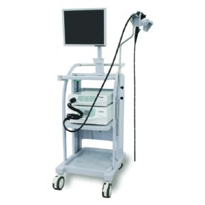
 AQ-100 equipped with high-resolution module (1280*1024) and CBI capabilities to provide the best possible image quality for endoscopes. Enhancing the observation of capillaries and mucosal issues. (HBE) Hemoglobin enhancement (CBI) Compound Band Imaging Fully digital AQ SYSTEM processor Electronic magniication (ENH) Outline enhancement
AOHUA Aq100 Imaging System
-
 To provide image processing functions and igure and blood vessels enhancement functions which increases e ciency of clinical diagnosis of diseases. Auto White balance adjustment function.Image brightness and color adjustment function. Images frozen and picture in picture function.Figure and Blood vessels enhancement function. Peak and average metering light Shift function. DVI Digital Signal Blood Vessels Enhancement 150W Xenon Lamp
AOHUA VME-2800 Imaging System
Zenrox Healthcare Solutions - Advanced Medical Equipment in Lagos
Zenrox offers cutting-edge medical equipment and comprehensive support services in Lagos. Explore our products designed for superior healthcare delivery. Contact us today for innovative medical solutions.