-
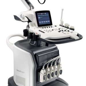
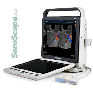
S40 Technology and Elegance 19 inch definition LCD monitor with wide viewing angle 10 inch touch screen with 15°adjustable angle Height and position adjustable control plain with muted function Five transducer sockets and additional one for CW probe Additional Endocavity probe holder and gel warmer Full range of transducers: Linear, Convex, Micro-convex, Endocavity, Phased array, Intraoperative, TEE, Pencil probe, Volumetric, Endocavity 4D and Laparoscope probe Advanced application technology: TDI, Stress Echo and Elastography Full patient database solution: DICOM 3.0, AVI/JPG, Dual USB, HDD, DVD, PDF report
Sonoscape S40 Ultrasound
-
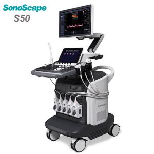
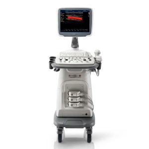
The design of s50 took operational use into consideration, creating a comfortable diagnosing enviroment. Ergonomic design, excellent man-machine interaction and rapid response, makes s50 an intelligent scanning assistant for you, bringing improved efficiency and helping to prevent fatigue from multiple examinations. 21.5 inches wide Led Monitor Super Responsive Touch Screen Flexible Adjusting Control Panel Built in Gel Warmer Wireless Wi-Fi Connection
Sonoscape S50 Ultrasound
-
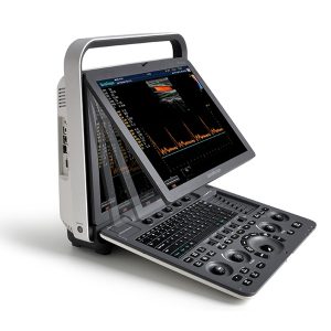
HotSonoscape S8 Exp Portable Ultrasound scanner DETAILS Agile and Versatile With ultra-modern innovative design and the clinically-proven technologies, S8 Exp Portable Ultrasound scanner is well equipped as a low-physical-effort and enhanced-image-quality ultrasound scanner, which not only provides optimized solutions for versatile applications but does help to improve the user experience for both routine and non-traditional challenges. Working with S8 Exp, it will trigger your unlimited reverie and endow you with endless charm. Carrying forward the classical design of SonoScape’s portable ultrasound products, S8 Exp successfully combines the best ergonomics, attractive design and ease of use. This charismatic identity is also enhanced by a sophisticated color palette—with sedate grey as its interior paint color and pearl white as exterior cover, S8 Exp reveals a style of aristocrat and strong character among SonoScape’s ultrasound systems. Workflow The S8 Exp is a portable ultrasound scanner that adapts to your workflow, whether you are in the consulting room, at the bedside, or at a remote location. With easy-to-use new platform designed for sonographers’ needs and full connection interfaces for easy connectivity and data sharing, S8 Exp leads to improved user comfort and clinical outcome as well as patient throughput and working efficiency. Powerful Platform Embedded with SonoScape‘s core imaging technologies such as μ-scan, PHI and Spatial Compound, S8 Exp boasts exceptional 2D image, sensitive spectral, Color and Power Doppler, displaying well-defined anatomy and pathology and facilitating a highly optimized diagnostic user environment for conclusive diagnoses. Besides, S8 Exp offers a comprehensive selection of electronic probes to maximally extend its capabilities to meet a wide range of applications including the abdomen, pediatric, OB/GYN, cardiovascular, musculoskeletal, etc. The advanced probe technologies also effectively enhance the image quality and confidence in reaching clinical diagnoses even in difficult patients. Click Here To Download Catalogue
Sonoscape S8 Exp Portable Ultrasound
-
LCD 15” Monitor angle adjustment 2 active probe connectors High density probes Scanning modes: В, М, В/М, В/В, 4В High precision tissue harmonics ZOOM at real time and frozen state Ultrasound tomography mode TRAPEZOIDAL mode at linear probes MicroScan speckle reduction imaging Color Doppler mode Power Doppler mode Directional power Doppler mode Pulse wave Doppler mode Continuous Doppler mode Color tissue Doppler mode Pulse wave tissue Doppler mode Duplex and Triplex modes Steer M-mode Color M-mode Free Hand 3D mode 4D mode PANORAMIC mode ELASTO with quantitative evaluation STRESS ECHO mode
Sonoscape S8EXP Ultrasound
-
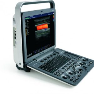
Modern technologies, innovative design and perfect imaging provide unlimited opportunities to the S9 system users LCD 15” Monitor angle adjustment 2 active probe connectors Touch control panel High density probes Scanning modes: В, М, В/М, В/В, 4В High precision tissue harmonics ZOOM at real time and frozen state Ultrasound tomography mode TRAPEZOIDAL mode at linear probes MicroScan speckle reduction imaging Color Doppler mode Power Doppler mode Directional power Doppler mode Pulse wave Doppler mode Continuous Doppler mode Color tissue Doppler mode Pulse wave tissue Doppler mode Duplex and Triplex modes Steer M-mode Steer M-mode Color M-mode Free Hand 3D mode 4D mode Panoramic Mode Elasto With Quantitative Evaluation Stress Echo Mode
Sonoscape S9 Ultrasound
-
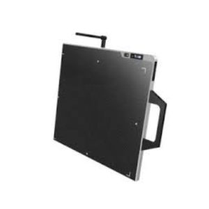
Features: Convenience: * AED (Automatic Exposure Detection) * Up to 3 detectors can be connected * Emergency Study * Optimized digital retrofit solution for X-ray system * Led lamp applied Speed: * Cycle time: less than 4.5 sec. * 1Gbps Ethernet * Steller gram, our console software, is simple, fast, stable and compatible * Promix: Signers processing tool * Client viewer (up to 5 places connectable, coming soon) Standard Accessories: Stellar M17 1Pc. DR: 17″x17″ Wired Detector CSI with Stellar Gram S/W 1Pc. Specification: Sensor: Amorphous Silicon Scintillator: CSI (Cesium-Iodide) Active Pixel Area: 427 x 427mm Pixel Pitch:139µm Resolution: 3.6 Ip/mm Pixel Matrix: 3072 x 3072 A/D Conversion:16 bit Cycle Time: <4.5 sec Interface (Wire):1Gbps Ethernet Dimension: 460mm(W) x 460mm(L) x 15mm(H) Weight: Detector: 4.0kg Console box: 1.1kg Operation Environment:50C to 350C, 30% to 85% RH Power: DC 24Vdc, 1.25A PC Min. Requirements: CUP: more than Intel Premium G5400 RAM: more than 8GB OS: Win 10 pro, 64bit Ethernet Card: Intel Gigabit Monitor: more than 17” Click Here To Download Catalogue
Stellar M17 CSI 17”x17” Wired Digital Radiography
-
Sale
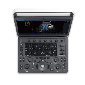
Ultrasound Machine – Sonoscape E2 – Color Ultrasound Scanner (Table Top)
Original price was: ₦13,800,000.00.₦13,437,500.00Current price is: ₦13,437,500.00.Original price was: ₦13,800,000.00.₦13,437,500.00Current price is: ₦13,437,500.00. Add to cartQuick View -
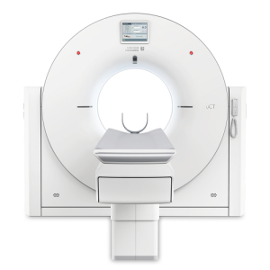
Technologies Single-click Bone Removal Employs different algorithms that are tailored to different applications such as carotid CTA, body CTA and virtual DSA. Single-click Rib Extraction Automatically segments the ribs and displays them separately or with the associated anatomy allowing efficient diagnosis of rib fractures. Highly Efficient Workflow EasyRegister The most frequently used protocols are placed in the front page shortcut area for easy selection. Along with the preset patient positions, the patient registration can be completed efficiently. EasyScan Intelligently plans the exam for individual patients. Clinical parameters are automatically adapted to the needs of the particular patient. EasyLogic The system anticipates the operator’s needs and prepares itself to reduce delays. Minimizes the wait time between operation procedures and accelerates CT exam workflow significantly. Innovative Z-Detector Fully Integrated Design Innovative Through-Silicon-Via (TSV) Technology. cm to μm level signal conducting path shortening. Ultra-low noise signal output. 0.55mm Pixel Size 0.5mm high resolution acquisition in all FOVs and collimations. 22mm coverage in Z-plane. 34560 large number of detector elements. Imaging with care KARL 3D Iterative Denoising The algorithm is performed in both the projection and image spaces to reduce dose while preserving image quality. 70kV Scan Mode 70kV Dose reduction can increase SNR for Iodine-based contrast agents especially for small BMI or younger patients. uDose 3D Dose Modulation Based on anatomic structure information, it generates optimized 3D dose distribution plan. Outstanding Financial Performance Reducing total cost of ownership for operation. Reliable Components Light-weight tube with reliable design and good serviceability. Self-cleaning carbon brushes for improved slip-ring reliability. Green Mode Switching the system into uECO mode during night or when the system is idle saves 30% power compared to conventional standby modes. Small Footprint uCT 520 requires a minimum installation area of 9m2. Advanced Applications Lung Nodule Analysis* A complete clinical solution including marking, segmentation and measurement assistance. Lung Density Analysis* Automatic lung extraction and contour editing with pulmonary density and volume measurement. CT Colonoscopy* Automatic segmentation of colorectum and electronic cleansing; virtual colonoscopy for a direct and efficient lesion measurement. Dental Analysis* Panoramic curve plotting, nerve line marking and sectional view analysis to diagnose dental diseases. * is optional. Clinical Gallery Head Inner Ear Lung Joint MAC (metal artifact correction) Lombar Spine Abdomen Body CTA
United Imaging CT Scan UCT 520
-
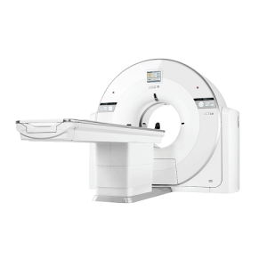
Technologies Z-Detector The Z-Detector reduces electronic noise down to a single photon level and the fully integrated design shortens the signal transmission length from centimeter to micron scale, resulting in tremendous electronic noise reduction and efficient X-ray utilization. KARL 3D Iterative Denoising The algorithm is performed in both the projection and image spaces to reduce dose while preserving image quality. Workflow with Ease EasyRegister The most frequently used protocols are placed in the front page shortcut area for easy selection. Along with the preset patient positions, the patient registration can be completed efficiently. EasyScan Intelligently plans the exam for individual patients. Parameters such as the scan range, reconstruction FOV and gantry tilt angle are automatically adjusted.* * Optional EasyLogic The system anticipates the operator’s needs and prepares itself to reduce delays. Minimizes the wait time between operation procedures and accelerates CT exam workflow significantly. Imaging with care 70kV Scan Mode 70kV Dose reduction can increase SNR for Iodine-based contrast agents especially for small BMI or younger patients. uDose 3D Dose Modulation Based on anatomic structure information, it generates optimized 3D dose distribution plan. Outstanding Financial Performance Reducing total cost of ownership for operation. Reliable Components Light-weight tube with reliable design and good serviceability. Self-cleaning carbon brushes for improved slip-ring reliability. Go Green Switching the system into uECO mode during night or when the system is idle saves 30% power compared to conventional standby modes. Small Footprint uCT 528 requires a minimum installation area of 9m2. Clinical Gallery uCT 528
United Imaging CT Scan UCT 528
-
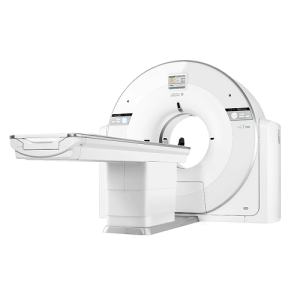
Technologies Ultra-Low-Noise Z-Detector The ultra-low noise detector fundamentally improves the image quality. With Through-Silicon-Via (TSV), Z-Detector enables direct output of digital signal, which achieves not only overall noise reduction, but also helps to ensure optimal image quality with low radiation dose. Iterative Denoising Technique The customizable KARL 3D iterative denoising algorithm maintains consistent image quality with a reduced dose. KARL 3D can also be combined with the metal artifact correction (MAC) algorithm to process scan data of patients with metal implants, eliminating metal artifacts and reducing image noise. Dedication to Fine Craftsmanship and Quality United Imaging has a remarkable dedication to craftsmanship and quality. From every detail inside the scanner to the finished product, the uCT 530 exudes fine craftsmanship and quality, which is evident in the stunning clinical images produced by the system. Highly Efficient Workflow High Throughput Hardware Platform The uCT 530 is equipped with a 5.3 MHU X-ray tube with fast heat dissipation capacity of up to 815 kHU/min, which ensures a fluid clinical workflow and offers high throughput. Easy-Logic Intelligent Prediction Platform The Easy-Logic intelligent prediction platform can predict the user’s next operation and initiate system preparation ahead of time. Factors such as tube preparation and early rotation of the gantry enable a seamless and efficient scanning process. uECO Energy-Saving Module The uECO shortens the time needed to stabilize the system after standby. The energy-saving module reduces the power consumption of the CT and HVAC systems. Poised. Proficient. Clinical Gallery Abdomen & Pelvis Study, Coronal View Carotid and Circle of Willis Study Brain uCT 530 BRAIN ABDOMEN & PELVIS STUDY, CORONAL VIEW CAROTID AND CIRCLE OF WILLIS STUDY BRAIN ABDOMEN & PELVIS STUDY, CORONAL VIEW CAROTID AND CIRCLE OF WILLIS STUDY BRAIN Passion For Change
United Imaging CT Scan UCT 530
-
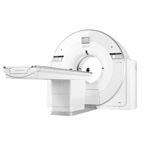
Technologies Z-Detector The Z-Detector, designed with a fully integrated architecture and powered by through-silicon via (TSV) technology, shortens the signal transmission path from cm-level to µm-level. The outstanding denoising performance substantially improves the signal-to-noise ratio (SNR) and significantly enhances the capability to capture fine details within images. Comprehensive Cardiac Imaging The Multi-Phase Cardiac Imaging Technique catches every heartbeat in order to provide accurate cardiac imaging. The integrated Vital Signal Module provides accurate and real-time acquisition of the ECG signal, as well as automated detection of arrhythmia and abnormality. Dedication to Fine Craftsmanship and Quality United Imaging has a remarkable dedication to craftsmanship and quality. From every detail inside the scanner to the finished product, the uCT 760 exudes fine craftsmanship and quality, which is evident in the stunning clinical images produced by the system. Low-Dose Scan Experience uDose Intelligent Tube Current Modulation The tube current is automatically adjusted to accommodate different anatomy and positions to avoid unnecessary radiation caused by overexposure. KARL 3D Iterative Denoising Technique KARL 3D employs a generalized gradient descent iterative de-noising technique in both the projection and image spaces. It achieves excellent image quality at low dose levels. Performance. Profound. Clinical Gallery Routine – Cerebral Infarction Low Dose – CCTA-80 kV Prospective Scan 0.9 mSv Cardiac – High BPM Applications – Vessel Analysis Lower Extremity CTA
United Imaging CT Scan UCT 760
-
Technologies Z-Detector The Z-Detector reduces electronic noise down to a single photon level, improving the signal-to-noise ratio (SNR) and providing more detailed images. The fully integrated design shortens the signal transmission length from centimeter to micron scale, resulting in tremendous electronic noise reduction and efficient X-ray utilization. Image Reconstruction The uCT 780 provides dedicated solutions for more image detail and faster acquisitions. With a 0.5 mm collimation, finer structures are better visualized with every scan. The 1024 x 1024 reconstruction matrix enhances the spatial resolution and helps reveal the smallest of details for the most challenging examinations. Dedication to Fine Craftsmanship and Quality United Imaging has a remarkable dedication to craftsmanship and quality. From every detail inside the scanner to the finished product, the uCT 780 exudes fine craftsmanship and quality, which is evident in the stunning clinical images produced by the system. Coordinated Control System Architecture Rapid Transfer of Massive Data The uCT 780 provides 360 million voxel data acquisition and reconstruction speed up to 60 IPS. System Hardware Synchronization System hardware synchronization offers precise radiation dose control and 0.3s high-speed gantry rotation. Easy-Logic Intelligent Prediction Platform The Easy-Logic intelligent prediction platform rapidly predicts the user’s next action and efficiently prepares the tube, generator, gantry, and data path in advance of the user’s action. Precision. Accelerated. Clinical Gallery Inner Ear 70kV Scan Mode Low Dose Cardiac Scan CABG Coronary Stent Large Range CTA
United Imaging CT Scan UCT 780
-
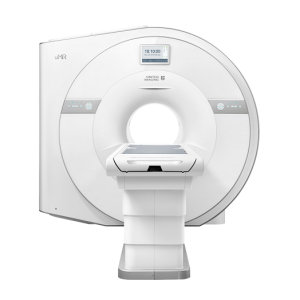
Technologies Full-Digital Imaging Chain The full-digital lossless RF waveform modulation technology is employed to achieve full-digital emission, transmission and reception of signals, so the image acquired is of high fidelity and low noise, resulting in superb image quality. High-Density Integrated Coils The combined imaging technology capturers signals of every coil element at the same time, providing imaging exams of multiple body parts with one scan. Intelligent System Management Automatic judgment of the system operation status; Fast wake-up in 5 seconds; Self-diagnosis of key functional components can greatly lower the operating cost of the system. Intelligent Early-Warning Real-time component monitoring is provided to improve system stability and reliability. Intelligent Wake-Up During the gap between exams, the system automatically enters the idle mode to save power. It can switch back to working mode in 5 secs if required. Intelligent Control Networks Intelligent and distributed system architecture design can offer an excellent system stability. The 16-channels RF system based on optical fiber transmission realizes low noise and distortion, presenting high fidelity images. Economic Design All core components are self-developed to ensure high system stability. Equipped with high quality, zero helium boil-off magnet, significant reduction in operation costs is realized. High Throughput Technologies such as full-digital RF system , high density integrated coil and unique bFAST parallel acquisition enable uMR 580 to significantly improve scanning efficiency and increase patient throughput. High System Stability The robustness and stability of the system is fully proven with 600,000 times bending tests of coils, 120,000 times plug tests of coil interface, 360,000 times travel tests of examining table, and full load operation test of gradient system. Zero Helium Boil-Off Extensive tests in clinical environments have demonstrated the magnet’s zero helium boil-off, producing a significant reduction in operation. Clinical Gallery Neuro Imaging Motion Artifact Reduction Non-contrast Angiography Imaging Fat Analysis & Calculation Technology High-resolution Musculoskeletal Imaging
United Imaging MRI UMR 580
-
uCS Imaging Technology Combing the strengths of PF, PI and CS, uCS (united Compressed Sensing) imaging technology has achieved optimized data acquisition and image reconstruction, significantly improving the scanning speed and image quality. uCS Application Achieving maximum acceleration factor of 16 for dynamic abdomen imaging with uCS, uMR 588 enables a clear visualization of every subtle and dynamic variations, and real-time lesion pinpointing in all orientations within the whole abdomen. uCS Imaging Driven by uCS imaging technology, uMR 588 breaks the limits of both resolution and speed of MRI. uExceed Workflow “Patient-oriented” Design uExceed software system enables parallel workflow patterns of multi-patient and multi-task, providing convenience for users and accelerating work efficiency. EasyScan Driven by EasyScan and whole body imaging, uMR 588 enables smart examination without moving patient table and changing coils manually, significantly simplifying the workflow and improving the throughout. Economic Success Intelligent System Management Monitor status of components in real-time; Intelligent analysis and forecast system trends; Timely service reminders Reduce Your TCO Zero helium boil-off; 10% energy saving; About 50% of liquid helium Clinical Gallery Neuro Spine Abdomen MSK Breast Angiography
United Imaging MRI UMR 588

Zenrox Healthcare Solutions - Advanced Medical Equipment in Lagos
Zenrox offers cutting-edge medical equipment and comprehensive support services in Lagos. Explore our products designed for superior healthcare delivery. Contact us today for innovative medical solutions.

 No products in the cart.