-
“Hersill Genesis Anaesthetic Machine” already exists in your wishlist
-
“ECG POCT” already exists in your wishlist
-
“Physio control LIFEPAK 1000 Defibrillator” already exists in your wishlist
-
“Philip AED Pad” already exists in your wishlist
-
“Maico MB11 Brainstem Evoked Response Audiometer” already exists in your wishlist
-
“Urit 12 Hemoglobin Meter” already exists in your wishlist
-
“Surgical Lights (Operating Lights)” already exists in your wishlist
-
“Hersill Genesis Anaesthetic Machine” already exists in your wishlist
-
“M30 Diluents” already exists in your wishlist
-
“Defibrillator - Philips” already exists in your wishlist
-
“Finecare Plus FIA Meter” already exists in your wishlist
-
“Mindray WATO A7 Anaesthesia Machines” already exists in your wishlist
“Hersill Genesis Anaesthetic Machine” was added to the compare list
“Physio control LIFEPAK 1000 Defibrillator” was added to the compare list
Compare“Ulna & Radius Dorsal Distal Radius Plate (Oblique)” has been added to the compare list
“Maico MB11 Brainstem Evoked Response Audiometer” was added to the compare list
“Philip AED Pad” was added to the compare list
View wishlist“Ulna & Radius Dorsal Distal Radius Plate (Oblique)” has been added to your wishlist
“ECG POCT” was added to the compare list
“Patient Monitor - Lerong” was added to the compare list
Compare“Timesco Ophthalmoscope” has been added to the compare list
View wishlist“Timesco Ophthalmoscope” has been added to your wishlist
View wishlist“Tibia & Fibula Reconstruction Plate” has been added to your wishlist
“Maico EasyTymp Screening Device” was added to the compare list
Compare“Tibia & Fibula Reconstruction Plate” has been added to the compare list
“Hersill Genesis Anaesthetic Machine” was added to the compare list
“ECG POCT” was added to the compare list
“M30 Diluents” has been added to your cart.
View cart
“M30 Diluents” was added to the compare list
“Finecare Plus FIA Meter” was added to the compare list
Compare“Portable Rebound Tonometer” has been added to the compare list
“Mindray WATO A7 Anaesthesia Machines” was added to the compare list
View wishlist“Portable Rebound Tonometer” has been added to your wishlist
“Mindray WATO EX-65/65 Pro Anesthesia Machines” was added to the compare list
“Maico TouchTymp MI36 Tympanometry” was added to the compare list
-

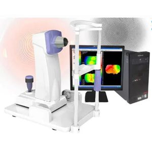
Corneal Topography Machine is a diagnostic tool that provides 3-D images of the cornea. The cornea is the outer layer of the eye, responsible for about 70 percent of the eye’s focusing power.
Features of Corneal Topography Machine:
1.By PLACIDO cone, 31 rings, a total of 7936 points.
2.Through calculation and analysis, used in the clinical diagnosis of corneal astigmatism, quantitative analysis of corneal shape, display the corneal curvature by data or different colors, can display axial curvature map, tangential curvature map, height map, simulated corneal lens map and corneal 3D map.
3.Can be used in preoperative examination and postoperative effect evaluation of corneal refractive surgery, can guide wearing contact data lens, to improve the accuracy of adaptive contact lens.
Technical Specifications of Corneal Topography Machine:
Model
Measuring method
Placido cone
Coverage
10.91mm(Diameter)
Curvature Radius
5.5mm-10.0mm
Diopter Range
33.75D-61.36D
Precision
±0.02mm
PlacidoRings
31 rings
Measurement Points
7936 points
Display
axial curvature map, tangential curvature map, elevation map, imitated keratoscope map and 3D cornea map
Image Output
High-quality color inkjet printer
Adjust range
left/right 86mm ; forward/backward 40mm ; up/down 30mm ; chinrest 5omm
Cornea contact lenses fitting function
With
Keratoconus detecting function
With
-

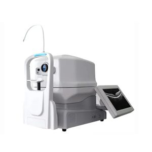
Optical Coherence Tomography Machine is an ophthalmic device for comprehensive eye exam, such as macular, retina problems or diseases, glaucoma, and so on. It is based on high sensitivity spectral domain technology to obtain high-resolution 3D retinal tomography images with high accuracy. It is fully automatic, simple and fast to operate. It provides several types of professional test reports to meet different examination requirements.
Features of Optical Coherence Tomography Machine:
1. The RetiView 500 provides a rich analysis software package with an automatic platform.
2. The RetiView 500 offers a special “one-key operation”mode which makes the captures simple and fast.
3. The RetiView 500 dramatically reduces the learning time and improves the success rate and your working efficiency.
4. The RetiView 500 has a compact design with a built-in computer which effectively saves space. The compact design also makes the shipment and movement easier.
5. The installation-free concept allows the immediate use of the device after connecting-the power cable and switching on the power.
6. The 13.3 inch touch screen allows easy operation with your finger and adjusting of the scanning parameters .
Specifications of Optical Coherence Tomography Machine:
Methodology
Spectral domain OCT
Axial resolution
≤ 6 µm (in tissue)
Transverse resolution
≤ 20 µm (in tissue)
Scan depth
≥ 2.5 mm (in air)
Scan range
≥ 6 mm
Scan speed
≥ 24,000 A-scans/sec, up to 36,000 A-scans/sec
Scan modes
30, Raster, Circle
Fundus image
OCT en face
Focus adjustment
-150 to 150
Pupil diameter
≥ 3 mm
OCT light source
840 nm SLD
Optical power
750 µW (at cornea)
Operation
13.3’’touch screen, optional external mouse or keyboard
Power supply
100-240 V, 50/60 Hz
Dimensions
497 mm × 395 mm × 490 mm (L× W × H)
Weight
LED dot matrix
External fixtion
34 kg (75 lbs)
-
Feature:
Large color liquid-crystal screen
Touch screen input, easy operation
Curve freezing: Manual/Auto mode, controlled by a pedal
Built-in speed thermal printer
Enter the name & ID; easy to check the archive
Technical Specifications:
Model
SW-1000 Ophthalmic A Scan Biometer
A Scan
A scan probe
10MHz import small size probe, built-in luminotron
Measuring range
15mm-40mm
Measurement precision
±0. 05mm; with macula lutea trace function
Measurement
Anterior chamber depth, lens thickness, vitreous body length, total length and average
Method of measurement
immersion and contact
Eye mode
Phakic/ Aphakic/Dense/ various IOL
IOL formula
SRK-II, SRK-T, BINKHORST- Ⅱ, HOLLADAY, HOFFER-Q, HAIGIS
Storage
10 cases, 5 readings each case
Output
A scan waveform and IOL calculation sheet
